
研究
【基本方針】
目指しています.
内容
1)
2) 定量的形態解析法の開発.
3)
ポリシー
FINER criteria (Cummings SR, et al 2001)
Novel(新しい知見を含む), Ethical(倫理上,問題がない),
Relevant
【業績】
 詳細へ
詳細へ
1)研究手法
a) プロトコール集
b) FISH法
組織細胞化学2006, 2008, 2011(FISHプロトコール)
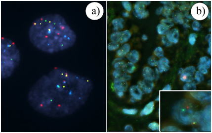
図: 染色体・遺伝子の数的異常と転座の検出(組織細胞化学2011,日本組織細胞化学会編).a) Multicolor FISH法.尿細胞診検体に対し,3番(赤), 7番(緑), 17番(青)染色体セントロメアと9p21領域(オレンジ色)に対するプローブ(UroVysion)を用いた.b) 分離シグナル検出法を用いた甲状腺乳頭癌におけるRET/PTC遺伝子転座の解析. RETの切断点を挟んで2つのBACプローブ(赤, RP11-124O1; 緑, RP11-168L22)を設計している.
b) Microscopic
FRET (Förster resonance energy transfer) 法
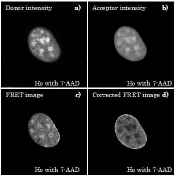
図;donorとacceptorの濃度により補正されたmulti-acceptor FRET画像(組織細胞化学2011,日本組織細胞化学会編).核内のHoechst 33258(Ho;
donor)と7-aminoactinomycin D(7-AAD;
acceptor)間で起こったFRET.
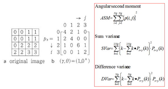
c) Texture analysis
濃度共起行列を用いた物質空間分布解析
2)診断病理学的研究
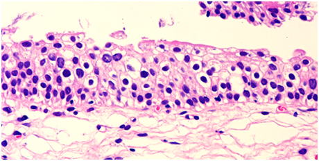
a)Unusual cytologic findings in low grade papillary transitional cell carcinoma.
(Acta Cytol).
図; G1相当の低異型度乳頭状尿路上皮癌の中には孤立散在性に腫瘍細胞が出現することがある.
b) Molecular and immunohistologic analyses cannot reliably solve diagnostic
variation of flat intraepithelial lesions of the urinary bladder. (Am J
Clin Pathol.)
図; 平坦状上皮内病変.反応性異型,異形成,上皮内癌と診断がばらついた症例.
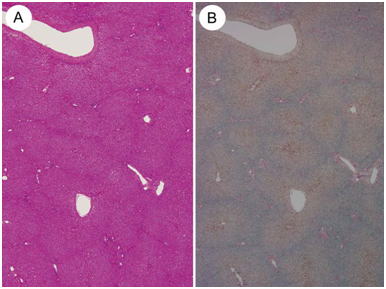
図 Focal nodular hyperplasiaの背景肝組織には鉄染色陽性の部分で囲まれる微小結節を認める.FNHの発生機序である血管異常との関連が示唆される.
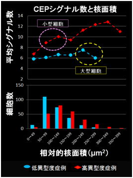
図; 低異型度および高異型度尿路上皮癌の細胞核面積と染色体・遺伝子異常の関係.高異型度症例は細胞が小型でも3, 7, 17番染色体の増加を伴っている.
3)定量的形態解析法の開発
a) Detection of underlying characteristics of nuclear chromatin patterns of
thyroid tumor cells using texture and factor analyses. (Cytometry)
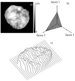
図; 甲状腺乳頭癌のTexture analysis. クロマチン分布の数値化.
b) Morphological abstraction of thyroid tumor cell nuclei using morphometry
with factor analysis. (Microsc Res Tech.)
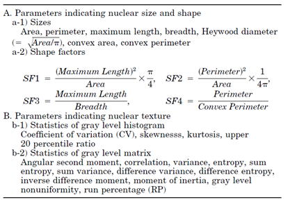
表; 細胞核形態(核形状およびクロマチン分布)を数値化するパラメータ.
c) Application of microscopic Forster resonance energy transfer to cytological
diagnosis of the thyroid tumors. (J Biomed Opt.)
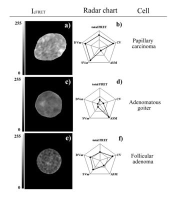
図; 甲状腺腫瘍性病変の細胞核内のFRET画像.Hoechst 33258と
4)腫瘍における構造異型および細胞異型の分子病理学的背景のresearch
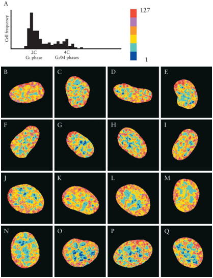
図: DNA histogram (A)と各細胞周期上の細胞核におけるHoechst 33258と
7-aminoactinomycin D間のcorrected
FRET画像.G1-phase (B–I), S-phase (J–M), and G2/M-phases
(N–Q)
b) Conservation and alteration of chromosome territory arrangements in thyroid carcinoma cell nuclei. (Thyroid.)

図; Chromosome territory.赤10番染色体,緑18番染色体,青19番染色体.
a) metaphase of lymphocyte, b) interphase of
lymphocyte, c) normal thyroid, d) nodular goiter, e) papillary carcinoma, f)
undifferentiated carcinoma.
c) Involvement of centrosomes in nuclear irregularity of thyroid carcinoma
cells (Virchows Arch)
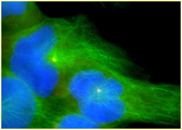
図; 甲状腺癌培養細胞に形成された核溝と核内細胞質封入体.緑;alpha-tubulin,黄色;gamma-tubulin
d) 甲状腺腫瘍細胞と核膜蛋白発現の異常
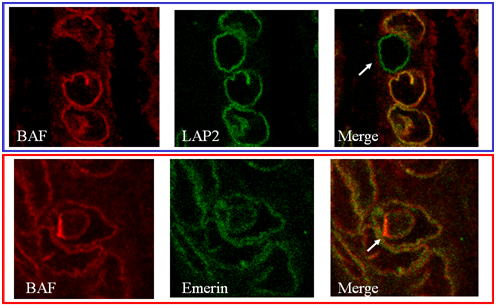
図: 甲状腺乳頭癌における核膜蛋白発現の異常
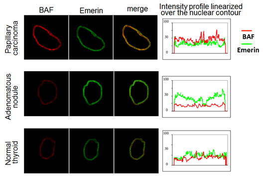
図: 甲状腺腫瘍細胞核上の核膜蛋白の分布異常
e) 発癌および化生におけるRNA 転写制御に関わる染色体高次構造の解析 (埼玉医科大学雑誌)
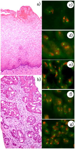
図; Subchromosomal positioning.1番染色体のchromosome territory(赤)とEpidermal
differentiation complex (EDC)遺伝子(緑)の関係.HE組織像(a, b)とmulticolor FISH像 (棘細胞 (c), 基底細胞 (d),腺細胞(e),扁平上皮化生細胞 (f)およびリンパ球(g).
和歌山県立医科大学人体病理学教室
〒641-8509
和歌山市紀三井寺811-1 アクセス
TEL 073-441-0635
FAX 073-444-5777
w-hupath@wakayama-med.ac.jp
サイト内検索